BIOLOGY Form 2 Topic 4
TRANSPORTATION OF MATERIALS IN LIVING THINGS
The Meaning of Osmosis, Diffusion and Mass- Flow
Explain the meaning of osmosis, diffusion and mass- flow
Life processes in organisms take place at the cell level. Therefore, it is necessary for substances to move in and out of the cells. There are two ways through which substances can move across the membrane. Materials in living organisms move by diffusion, osmosis and mass flow.
Diffusion
This is the movement of materials from a region of higher concentration to a region of lower concentration until equilibrium of two sides is maintained. Diffusion can also be defined as the movement of ions or molecules from the region of higher concentration to the region of lower concentration, without involving any permeable membrane. A difference in concentration of a substance between two regions is known as concentration gradient.
Diagram showing diffusion
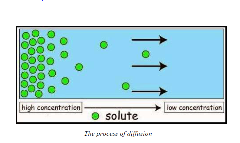
Materials are transported in the body system of living things from the area where they are abundant to areas where they are less abundant, and this process or mechanism of transportation in these animals is termed as diffusion. Diffusion occurs in exchange of gases like oxygen or carbon dioxide during respiration in animals and plants. Also, diffusion takes place during distribution of nutrients and digested foods in living organisms.
Osmosis
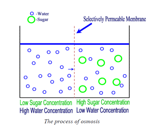
This is the movement of water molecules from a region of higher concentration to a region of lower concentration through a semi-permeable membrane. A partially-permeable membrane is a membrane that allows small particles such as water molecules to pass through it, but not larger particles such as sugar molecules and ions from salts. Examples of semi-permeable membranes are cell membranes and a pig’s bladder. These membranes allow transportation of water through them. In spite of the fact that they allow transportation of water through them, they do not permit the passage of sugar or salt molecules because they are solutes. Osmosis occurs when water moves down its concentration gradient across the semi-permeable membrane.
Therefore, for osmosis to take place there must be:
- two solutions with different concentrations; and
- a partially permeable membrane to separate them.
A dilute solution has a high water concentration, while a concentrated solution has a low waterconcentration. For example, when salt is dissolved in water:
- A little dissolved salt produces a dilute solution with a high water concentration
- A lot of dissolved salt produces a concentrated solution with a low water concentration.
Diagram of osmosis
Mass flow
Diffusion and osmosis occurs very slowly and cover short distances. In animals and plants,materials are usually transported a long distance and in large quantities. For example, food nutrients from the small intestine have to be moved to cells in the extremities such as toes and fingers, where the nutrient materials have to be transported a long distance. Therefore, an efficient and fast mechanism is required to facilitate this movement. That is when mass flow comes in.
Mass flow is the movement of materials in large quantities and across a long distance in the body of an organism due to differences in pressure between the two regions. Materials in higher plants and animals are moved by the process of mass flow. For example, the manufactured food in plant leaves has to be moved to all plant parts, for use or storage, by mass flow.
Experiments to Demonstrate the Process of Diffusion, Osmosis and Mass Flow
Carryout experiments to demonstrate the process of diffusion, osmosis and mass flow
Demonstration of the process of diffusion
Take a bottle of perfume and move to one corner of the classroom. Open a bottle and observe what happens. The result is, after a few seconds, the whole classroom is filled with a smell of the perfume. This means that the molecules of the perfume moves from the region of higher concentration (the bottle) to a region of lower concentration (air). That is why the smell is felt by a person standing several meters away from the source of the perfume.
Some important processes that involve diffusion are:
- Gaseous exchange in the lungs of animals and in the leaves of plants
- Absorption of digested food in the ileumThe process of diffusion
- Removal of west materials from cells
- Absorption of nutrients and oxygen into cells
Demonstration of the process of osmosis
Procedure
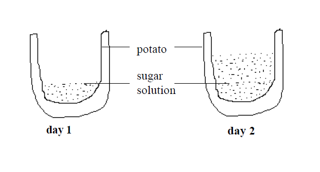
Peel a potato and cut it as shown in the diagram below. Then fill the depression with brine(concentrated solution of sodium chloride). Leave the set up until the next day and observe what happens to the level of brine in the potato.
Result
In the following day, you will find that the level of brine will have risen as shown. This means that water has moved from the potato to the brine solution causing the brine level to rise up. The water has moved from a region of high water concentration (the potato) through the cell membranes of the potato cells (partially permeable membrane) to the region of low water concentration (the brine).
The Differences between Diffusion, Osmosis and Mass Flow
Outline the differences between diffusion, osmosis and mass flow
Differences between diffusion and osmosis
| Diffusion | Osmosis |
| It is the movement of all types of substancesfrom the area of their higher concentration tothe area of their lower concentration | It is the movement of only solvent or waterfrom the area of their higher concentration tothe area of their lower concentration through apartially permeable membrane |
| Diffusion can operate in any medium | Osmosis operates only in a liquid medium |
| Diffusion is applicable to all types ofsubsstances (soilds, liquids and gases) | It is applicable only to solvent part of asolution |
| It does not require any semi-permeablemembrane | A semi-permeable membrane is a must foroperation of osmosis |
| It is purely dependent upon the free energy ofthe diffusing substance | Osmosis is dependent upon the dregree ofreduction of free energy of one solvent overthat of another |
| It helps in equalizing the concentration of thediffusing substance througout the availablespace | It does not equalize the concentration ofsolvent on the two sides of the system |
| Turgor pressure or hydrostatic pressure doesnot normally operate in diffusion | Osmosis is oppossed by turgor or hydrostaticpressure of system |
| It is not influenced by solute potential | Osmosis is dependent upon the solute potential |
| Diffusion of a substance is mostly dependentof the presence of other sustances | It is dependent upon the number of particles ofother substances dissolved in a liquid |
| Factors like water potential, solute potentialand pressure potential do not affect diffusion | Factors like water potential, solute potentialand pressure potential affect osmosis in aliving system |
The Roles of Diffusion, Osmosis and Mass Flow in Movement of Materials in Living Organisms
Explain the roles of diffusion, osmosis and mass flow in movement of materials in living organisms
Materials are transported in the body system of living things from the area where they areabundant to areas where they are less abundant, and this process or mechanism of transportationin these animals is termed as diffusion. Diffusion occurs in exchange of gases like oxygen orcarbon dioxide during respiration in animals and plants. Also, diffusion takes place duringdistribution of nutrients and digested foods in living organisms.
- Through the process of osmosis, nutrients get transported to cells and waste materials getmoved out of them.
- The pressure within and outside each cell is maintained by osmosis as this process ensures abalance of fluid volume on both sides of the cell wall. If fluid volume within a cell is morethan the fluid volume outside it, such pressure could lead the cell to become turgid andexplode. On the contrary, if fluid volume outside the cell is more than the fluid volumewithin, such pressure could lead the cell to cave in. Both cases would be detrimental tonormal and healthy cellular function.
- It is via osmosis only that roots of plants are able to absorb moisture from the soil andtransport it upwards, towards the leaves to carry out photosynthesis. Plants wouldn’t existwithout osmosis; and without plants, no other life could exist as they are a vital link of theentire food chain of the planet.
- Without osmosis, it would be impossible for our bodies to separate and expel toxic wastesand keep the bloodstream free from impurities. The process of blood purification is carriedout by the kidneys which isolate the impurities in the form of urine.
- Therefore, the role of osmosis is twofold: it helps maintain a stable internal environment in aliving organism by keeping the pressure of intercellular and intracellular fluids balanced. It alsoallows the absorption of nutrients and expulsion of waste from various bodily organs on thecellular level. These are two of the most essential functions that a living organism cannot dowithout.
The External and Internal Structures of the Mammalian Heart
Describe the external and internal structures of the mammalian heart
TRANSPORT OF MATERIALS IN MAMMALS
Mammals are the complex multicellular organisms whose bodies are made up of numerous cellsand tissues. In this case, diffusion alone is not enough to insure efficient carrying out of lifeprocess. Therefore mammals have an elaborate transport system that is made up of the heart,blood and blood vessel.
The structure of the mammalian heart
The heart is a muscular organ about the size of a closed fist that functions as the body’scirculatory pump. It takes in deoxygenated blood through the veins and delivers it to the lungsfor oxygenation before pumping it into the various arteries. The heart is located in the thoraciccavity between the two lungs.
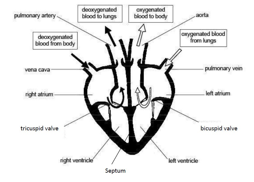
The external structure of the mammalian heart is as shown in the labelled diagram below:

The mammalian heart is broader at the top and narrower at the bottom. It is enclosed by a double layer of tough and elastic membranes called pericardium. These membranes prevent the heart from ever-expanding when beating very fast. Also the pericardium secrets a fluid which enables the membranes to move smoothly against each other.
The walls of the ventricles are thicker than those of the auricles because the ventricles pumpblood a greater distance than the auricles. Auricles pump blood to the ventricles while theventricles pump blood to the other parts of the body.
The left ventricle is thicker than the right ventricle because the right ventricle pumps blood to thelungs while the left ventricle pumps blood to the rest of the body parts.The heart consists of four chambers, right and left atria and right and left ventricles. Thefunctions of each part and the associated structures are as follows:
- The right atrium links to the right ventricle by the tricuspid valve. This valve preventsbackflow of the blood into the atrium above, when the ventricle contracts.
- The left atrium links to the left ventricle by the bicuspid valve. This valve also preventsbackflow of the blood into the atrium above, when the ventricle contracts.
- Semi-lunar (pocket) valves are found in the blood vessels leaving the heart (pulmonary arteryand aorta). They only allow exit of blood from the heart through these vessels followingventricular contractions.
- Ventricles have thicker muscular walls than atria. When each atrium contracts, it only needsto propel the blood a short distance into each ventricle while ventricles pump blood to distantbody parts.
- The left ventricle has even thicker muscular walls than the right ventricle. The left ventricleneeds a more powerful contraction to propel blood to the systemic circulation (all of the bodyapart from the lungs). The right ventricle propels blood to the nearby lungs. So, thecontraction does not need to be so powerful.
INTERNAL STRUCTURE OF THE MAMMALIAN HEART
The heart has several valves. And these valves have flaps that ensure that blood flows in one direction only.
These valves include the following:
- The tricuspid valve; found between the right auricle and right ventricle
- The bicuspid valve: found between left auricle and left ventricle
- Semi-lunar valves which are located at the bases of the pulmonary artery and aorta to preventblood from flowing back into the ventricles.
These valves will close if the blood flows back. The valves are held in place by tendons which prevent the flaps from turning inside out.The right and left sides of the heart are separated by septum which is a thick muscular wall which prevents mixing of oxygenated and deoxygenated blood.
The Functions of the External and Internal Parts of the Mammalian Heart
Explain the functions of the external and internal parts of the mammalian heart
Functions of parts of the mammalian heart
| Part of the heart | Function |
| Aorta | The largest artery in the body; it conducts freshly oxygenated bloodfrom the heart to the tissues. |
| Superior vena cava | Large vein that brings deoxygenated blood from the upper parts ofthe body to the right atrium |
| Inferior vena cava | Large vein that brings deoxygenated blood from lower regions of thebody to right atrium |
| Pulmonary artery | Carries deoxygenated blood from the right ventricle to the lungs |
| Pulmonary vein | Blood vessel that carries oxygenated blood from the lungs to the leftatrium |
| Right atrium | This chamber of the heart receives deoxygenated blood from thebody (from the superior and inferior vena cava). |
| Left atrium | This chamber of the heart receives oxygenated blood from the lungs |
| Tricuspid valve | Located on the right side of the heart between the right atrium (RA)and right ventricle (RV) |
| Bicuspid valve | Located on the left side of the heart between the left atrium (LA) andthe left ventricle (LV) |
| Right ventricle | The chamber of the heart that pumps deoxygenated blood to thelungs |
| Left ventricle | Receives blood from the left atrium and pumps it into the aorta fortransport to the body cells |
| Septum | Divides the right and left chambers of the heart |
The Adaptations of the Parts of the Mammalian Heart to their Functions
Explain the adaptations of the parts of the mammalian heart to their functions
The heart is adapted to carry out its functions by having the following features:
- The cardiac muscle is adapted to be highly resistant to fatigue.
- The heart has a large number of mitochondria enabling continuous supply of energy to theheart and numerous myoglobins (oxygen storing pigment).
- The presence of the cardiac muscles enables the heart to beat rhythmically.
- The pericardium which surrounds and protects the heart from physical damage.
- Pericardial fluid which prevents friction when the heart beats.
- The outer layer of the pericardium attaches to the breastbone and other structures in the chestcavity and thus helps to hold the heart in place.
- Bicuspid and tricuspid valves between atria and ventricles which prevent the backflow ofblood.
- Septum which prevents the mixing of deoxygenated blood in the right and oxygenated bloodin the left chambers of the heart.
- Its own blood supply for supplying nutrients and removing waste.
- The left ventricle has thick muscular wall to pump blood at a higher pressure to the distantbody tissues,
- The heart is supplied with the nerves which control the rate of heartbeat depending on thebody requirements.
Blood vessels
Blood vessels are intricate networks of hollow tubes that transport blood throughout the entirebody. This is an essential function as blood delivers valuable nutrients to and removes wastesfrom our cells. Blood vessels are constructed of layers of connective tissue and muscle. Theinner blood vessel layer is formed of endothelium. In capillaries and sinusoids, endotheliumcomprises the majority of the vessel. There are three types of blood vessels namely arteries,veins and capillaries. Each of these vessels has a different structure and function.
The Structure of Arteries, Veins and Capillaries
Describe the structure of arteries, veins and capillaries
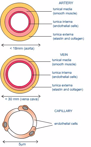
Basic structure
- Capillaries consist of anendothelium whichis only one cell thick.
- Walls of arteries and veins consist of 3 layers.
- The inner layer consists of a thin layer of endothelial cells.
- The middle layer is made up of smooth muscle with some elastic fibres. This layer controls the diameter of the vessel and hence the amount of blood and its rate of flow.
- The outer layer is composed of connective tissue; this holds the blood vessels in place in the body.
Detailed structure
Arteries
- The walls of arteries are much thicker as it carries blood away from the heart at high pressure.
- Major arteries close to the heart also have thick layers of smooth muscle in their walls to withstand the increases in pressure as the heart pumps.
- The walls also have a large proportion of elastic fibres in both the inner and middle layers – this allows for the arteries to stretch according to the increases in volume of blood. As the heart relaxes the artery walls return to their original position, hence pushing the blood along – maintaining a constant flow in one direction.
- Arteries are near the surface of the skin; the changes in the arteries diameter can be felt as a pulse.
Veins
- The walls of veins are thinner than the walls of arteries, as the blood they receive from the capillaries is at a much lower pressure.
- The walls have fewer elastic fibres and the lumen is wider (to allow for easier blood flow).
- Veins have two mechanisms for keeping the blood flow constant and in one direction. Firstly, many veins are close to muscles, hence when the muscles contract they compress the walls of the vein – pumping blood forwards. Veins also havevalves which are spacedalong regular intervals in veins. They work much like one-way swinging doors – as the blood is forced through the valve opens. However, once the pressure drops and the blood flow decreases, the valve shuts – preventing backflow of blood.
Capillaries
- They are extremely, tiny microscopic vessels that bring blood into close contact with the tissues, for the exchange of chemical substances between cells and the bloodstream.
- The one cell thick endothelial layer is a continuation of the lumen arteries and veins.
- Diffusion is a relatively slow process and hence the structure of capillaries is suited to slowing down the flow of blood.
- In order to maximize the exchange of substances between the blood and cells, capillaries have thin walls (for more efficient diffusion) a small lumen (that forces blood cells to pass through in single file, slowing down the rate of flow and maximizing their exposed surface area).
- They form an expansive blood flow network, such that no cells are far from blood supply
How different blood vessels are adapted for their function
| Blood vessel | Function | Adaptation |
| Artery | Carries blood away from heart at high pressure | Thick, elastic, muscular walls to withstand pressure and to exert force (pulse) |
| Vein | Returns low-pressure blood to heart | Large diameter to offer least flow resistance. Valves to prevent back flow. |
| Capillary | Allows exchange of materials between blood and tissues | Thin, permeable walls |
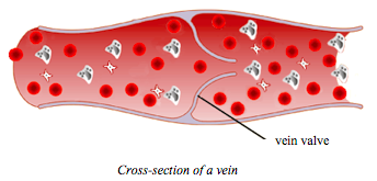
Cross section of vein
Differences between arteries, veins and capillaries
| Arteries | Veins | Capillaries |
| All arteries carry bloodawayfrom the heart | All veins carry bloodtowardsthe heart | Capillaries carry bloodfrom arteries to the veins |
| With the exception of the pulmonary artery, all arteries carryoxygenatedblood | With the exception of the pulmonary vein, all veins carrydeoxygenatedblood | Bloodslowly loses its oxygen |
| They carry blood which is usuallyrich in digested foodmaterials | Except for the hepatic portal vein, they carry blood which usuallyhaslittledigested foodmaterials | Bloodslowly loses its food |
| Have relatively narrower lumens (see diagrams above) | Have relatively wide lumens (see diagrams above) | Have relatively narrow lumens (see diagrams above) |
| Have relatively athicklayer of muscles and elastic fibres | Have relatively athinlayer of muscles and elastic fibres | Theydo not havemuscles and elastic fibres |
| They havethick outer walls | They havethin outer walls | Walls are onlyone cell thick |
| They carry blood athigh pressure | They carry blood atlow pressure | Pressure gradually fallsas blood flows from arteries to veins |
| Do not have valves (except for the semi-lunar valves of the pulmonary artery and the aorta) | Have valves throughout the main veins of the body to prevent the back flow of blood. | Have no valves |
| Have bright red blood (because it is rich in oxygen) | Brown-red blood | Brown-red blood |
| Located deep in the to body surface | Located near to body surface | Capillaries are found inside all tissues |
| Walls arenot permeable | Walls arenot permeable | Walls arepermeable |
| Blood flows in pulses | Nopulse | Pulse gradually disappears |
Simple Experiments to Determine Pulse Rates in Human Being
Carry out simple experiments to determine pulse rates in human being
Activity 1
Carry out simple experiments to determine pulse rates in human being
The Major Components of the Blood
List the major components of the blood
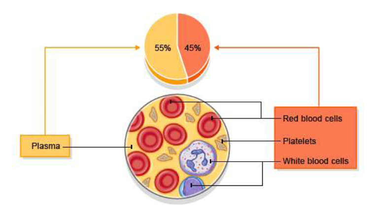
Blood is the red fluid that circulates in our blood vessels. The average human body containsabout 4 to 5 litres of blood. Blood is classified as a connective tissue and consists of two maincomponents:
- Plasma which is a clear extracellular fluid.
- The solid component, which are made up of the blood cells and platelets
The solid component is made up of blood cells except for the platelets, which are tiny fragmentsof bone marrow cells.
The solid component consists of blood cells (corpuscles) which include:
- Erythrocytes, also known as red blood cells (RBCs)
- Leukocytes, also known as white blood cells (WBCs)
- Platelets, also known as thrombocytes
Red blood cells, most white blood cells, and platelets are produced in the bone marrow, the softfatty tissue inside bone cavities. The white blood cells (lymphocytes) are also produced in thelymph nodes and spleen, and in the thymus gland.
Within the bone marrow, all blood cells originate from a single type of unspecialized cell calleda stem cell. When a stem cell divides, it first becomes an immature red blood cell, white bloodcell, or platelet-producing cell. The immature cell then divides, matures further, and ultimatelybecomes a mature red blood cell, white blood cell, or platelet.
Blood cells

By volume, the plasma constitutes about 55% of whole blood, and red blood cells, platelets and white blood cells about 45%.
The Function of Major Blood Components
Explain the function of major blood components
Red blood cells
Red blood cells (RBCs) have two main functions:
- To pick up oxygen from the lungs and deliver it to tissues elsewhere.
- To pick up carbon dioxide from other tissues and unload it in the lungs.
Erythrocytes transport oxygen in the blood through the red pigment called haemoglobin.Haemoglobin contains iron and proteins joined to greatly increase the oxygen carrying capacityof erythrocytes. The high surface area to volume ratio of erythrocytes allows oxygen to be easilytransferred into the cells in the lungs and out of the cells in the capillaries of the systemic tissues.Erythrocytes are produced inside red bone marrow from stem cells at the astonishing rate ofabout 2 million cells every second.
White blood cells
Although the white blood cells accounts for only about 1% of the blood, they play a veryimportant role in the body. Their main function is to protect the body against disease pathogens.There are two of white blood cells, each of which plays a specific role in protection of the bodyagainst illness and disease.
- Phagocytes: Engulf and digest invading bacteria and viruses (pathogens). It is the body’smain defence against germs (microbes).
- Lymphocytes: produce antibodies which neutralize antigens from bacteria or viruses. Theykill microbes or make them clump together, to be removed in the lymph glands.
White blood cells are produced in the yellow marrow of the bone, spleen, thymus and lymphaticsystem.
Platelets
Platelets are small fragments of bone marrow cells and are therefore not really classified as cellsthemselves. Platelets have the following functions:
- Secrete vasoconstrictors which constrict blood vessels, causing vascular spasms in brokenblood vessels.
- Form temporary platelet plugs to stop bleeding.
- Secrete procoagulants (clotting factors) to promote blood clotting.
- Dissolve blood clots when they are no longer needed.
- Digest and destroy bacteria.
- Secrete chemicals that attract neutrophils and monocytes to sites of inflammation.
- Secrete growth factors to maintain the linings of blood vessels.
In general, the blood platelets functions in healing of the wounds when the skin gets broken. Thisis achieved by clumping together of the platelets to form a network of mesh, hence bleeding isstopped.
Plasma
Plasma is the non-cellular or liquid portion of the blood. Plasma is a mixture of water, proteins,and dissolved substances. Around 90% of plasma is made of water, although the exactpercentage varies depending upon the hydration levels of the individual. Blood plasma has thefollowing functions:
- Plasma serves as a transport medium for delivering nutrients to the cells of the various organsof the body.
- It transports waste products derived from cellular metabolism to the kidneys, liver, and lungsfor excretion.
- It fights infections since it contains antibodies.
- It is also a transport system for blood cells, and it plays a critical role in maintaining normalblood pressure.
- Plasma helps to distribute heat throughout the body and to maintain homeostasis, orbiological stability, including acid-base balance in the blood and body.6. It carries and transports some hormones.
The Effects of HIV on White Blood Cells
Explain the effects of HIV on white blood cells
The HIVs in the blood of a HIV-positive person attack the white blood cells (lymphocytes). Theviruses reproduce and increase in number within the lymphocytes. Then the lymphocytes burstand release more viruses in the bloodstream. The released viruses attack more, new white cells.The attack continues in that cycle until many white cells are destroyed. Because there are only afew white cells left to fight against pathogens, the body immunity gets low. Once the immunityis lowered, the body is often attacked by diseases and a person suffers from AIDS.
The Concepts of Blood Group and Blood Transfusion
Explain the concepts of blood group and blood transfusion
Human blood can be grouped into four blood groups namely groups A, B, AB and O. They werediscovered in 1900 and 1901 at the University of Vienna by Karl Landsteiner in the process oftrying to learn why blood transfusions sometimes cause death and at other times save a patient.This classification is based on the type of antigens in the red blood cells and antibodies in theplasma.
Red blood cells have proteins (antigens) on their surface: A, B or A and B. Plasma hasantibodies which can cause agglutination: anti-A and anti-B.
Serum is blood plasma without fibrinogen. It can be stored without clotting, and is used intransfusions.
| Blood group | Antigen | Antibodies | Agglutinates |
| A | A | Anti-B | Anti-A serum |
| B | B | Anti-A | Anti-B serum |
| AB | A and B | None | Anti-A and anti-B serums |
| O | None | Anti-A and anti-B | Neither serum |
Consider the table above. People with type A blood will have the A antigen on the surface oftheir red cells (as shown in the table). As a result, anti-A antibodies will not be produced bythem because they would cause the destruction of their own blood. However, if B type blood isinjected into their systems, anti-B antibodies in their plasma will recognize it as alien and burstor agglutinate the introduced red cells in order to cleanse the blood of alien protein.
Individuals with type O blood do not produce any antigens. Therefore, their blood normally willnot be rejected when it is given to others with different blood types. As a result, type O peopleare universal donors for transfusions, but they can receive only type O blood themselves. Thosewho have type AB blood do not make any antibodies. Their blood does not discriminate againstany other blood type. Consequently, they are universal receivers for transfusions, but their bloodwill be agglutinated when given to people with every other type because they produce both kindsof antigens.
Blood grouping
It is easy and inexpensive to determine an individual’s blood type from a few drops of blood.This is how blood typing/grouping it is done: A serum containing anti-A antibodies is mixedwith some of the blood. Another serum with anti-B antibodies is mixed with the remainingsample. Whether or not agglutination occurs in either sample indicates the blood type. Forinstance, if an individual’s blood sample is agglutinated by the anti-A antibody, but not the anti-Bantibody, it means that the A antigen is present but not the B antigen. Therefore, the blood typeis A.
Rhesus factor
Some people have another antigen called Rhesus antigen on their red blood cells while othersdo not have it. Those having this antigen are referred to as Rhesus positive (Rh+) and thosewithout are it are Rhesus negative (Rh-). Rh antigen occurs in red blood cells and Rh antibodyoccurs in blood plasma.
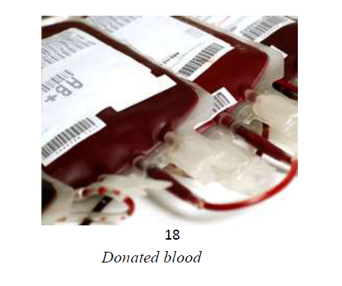
If Rh antibody mixes with Rh antigen during blood transfusion, agglutination will occur. Rh+ canstimulate the Rh- to produce antibodies to act against Rh+ antigens. However, the Rh- cannotstimulate the Rh+ blood to produce antibodies against Rh-. Therefore, an Rh+ person can receiveblood from the Rh- donor. The donated blood below is group AB rhesus positive (AB+).
The Relationship between Blood Groups and Blood Transfusion
Outline the relationship between blood groups and blood transfusion
Blood transfusion
Blood transfusion is the transfer of blood from one person (donor) to another person (recipient)through blood vessels. Transfusion is done to replace lost blood due to illness, accidents or bleeding. The donor is the person who gives blood while the recipient is the person who receives blood.
When performing blood transfusions it is important to avoid combining corresponding antigensand antibodies because they cause agglutination of red blood cells which may lead to death ofthe recipient. Agglutination is the clumping of red blood cells. Blood transfusion is only possibleif blood groups are compatible. Blood group compatibilities are as shown in the table below.
| Recipient | Donor | |||
| A | B | AB | O | |
| A | √ | × | × | √ |
| B | × | √ | × | √ |
| AB | √ | √ | √ | √ |
| O | × | × | × | √ |
Note: a tick (√) means compatible and a cross (×) means incompatible.
Individuals with blood group AB can receive blood from individuals of all blood groups and areknown as universal recipients. Individuals with blood group O can donate blood to individualsof all blood groups and are known as universal donors.
The Advantages and Disadvantages for Blood Transfusion
Explain the advantages and disadvantages for blood transfusion
Advantages of blood transfusions
Blood transfusion does so much for patients in need. The gift of life is donated, tested, processedand sent to hospitals’ transfusion service departments where more important work is done toensure it is compatible with the recipient.
Blood transfusion has a number of advantages. These are some of the benefits the donated bloodcan provide for patients in need:
- Increase low haemoglobin levels: low haemoglobin can cause damage to body organs andtissues due to low oxygen levels. Donated blood, with sufficient haemoglobin, can correct theproblem of low haemoglobin level of the recipient.
- Help stop bleeding: bleeding may not be controlled if platelets and/or clotting factors arelow. Receiving blood with high clotting factors can solve the problem.
- Keeps the heart pumping: low blood volume can lead to low pressure and the heart may notbe able maintain the circulation of blood.
- Help with serious blood infections when other methods fail. For example, blood transfusionmay serve as a treatment method for people with sickle cell anaemia or blood cancer(leukaemia).
- Provide red cells and platelets when the bone marrow is compromised as with blood cancers,bone marrow transplants, chemotherapy, etc.
- Provide red cells and platelets for patients with blood disorders such as sickle cell.
- Save someone’s life: people who have had a big loss of blood due to a number of reasons canhave their lives saved once they receive donated blood.
- Because blood transfusion involves screening of the donor’s blood, if the donor has anyhealth problem it can be detected and hence treated before getting worse.
Disadvantages of blood transfusions
Although blood transfusions can be life-saving, they are not without risks. The following aredisadvantages of blood transfusions:
Medical reactions:
- Allergic reaction: This is the most common reaction. It happens during the transfusionwhen the body reacts to plasma proteins or other substances in the donated blood.
- Fever reaction: The person gets a sudden fever during or within 24 hours of thetransfusion. Headache, nausea, chills, or a general feeling of discomfort may come withthe fever.
- Haemolytic reactions: In very rare cases, the patient’s blood destroys the donor red bloodcells. This is called haemolysis. This can be severe and may result in bleeding and inkidney failure.
Diseases:If proper screening of the donated blood is not observed, it can causetransmission of diseases from the donor to the recipient. Examples of such transmissiblediseases are HIV virus, hepatitis, and other infections.
Patients who are given too much blood can develop high blood pressure, a concern forpeople who have heart disease.
Precautions to be Taken During Blood Transfusion
Outline precautions to be taken during blood transfusion
Blood transfusion precautions
Certain precautions and guidelines must be adhered to in blood transfusion to ensure the safetyof the procedure. The precautions may include the following:
- Donated blood must carefully and thoroughly be screened for any infectious diseasesbefore being transfused to the recipient. The blood should be screened for diseases likehepatitis B, HIV virus, and all sexually transmitted diseases (STDs).
- The donated blood must be matched with the recipient’s blood type, as incompatibleblood types can cause a serious adverse reaction (transfusion reaction). Blood isintroduced slowly by gravity flow directly into the veins (intravenous infusion) so thatmedical personnel can observe the patient for signs of adverse reactions.
- During blood transfusion, vital signs such as body temperature, heart rate, and bloodpressure are carefully monitored.
- Some patients may get a sudden fever during or within 24 hours of the transfusion, whichmay be relieved with pain-relieving drugs such as panadol, diclofenac or paracetamol.This fever is a common reaction to the white blood cells present in donated blood.
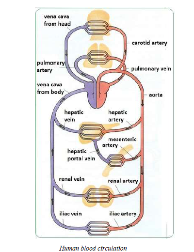
Blood Circulation in Humans
Describe blood circulation in humans
Blood circulation is the flow of blood from the heart to all body parts and back to the heart.Blood circulation or circulatory system, also called cardiovascular system, is one of three mainsystems in human body which consist of organs and tissues.
The cardiovascular systems of humans are closed, so the blood never leaves the network of bloodvessels. But oxygen and nutrients diffuse across blood vessel layers and enter interstitial fluid,which carries it to the target cells and carbon dioxide and wastes in the opposite direction.
The human blood circulation consists of two circulations namely the pulmonary circulation andsystemic circulation.
Pulmonary circulation
Pulmonary circulation is the movement of blood from the heart, to the lungs, and back to theheart again. This is just one phase of the overall circulatory system. In this type of circulation,the blood flows from the right ventricle to the lungs and from the lungs to the left auricle. In thepulmonary circulation, the blood circulates to and from the lungs, to release the carbon dioxideand pick up new oxygen.
In the pulmonary circulation, blood from all body parts (except the lungs) enters the right auriclethrough vena cava. From the right auricle the blood descends into the right ventricle through thetricuspid valve. When the ventricle contracts, the blood is pushed into the pulmonary artery thatbranches into two main parts: one going to the left lung, and another to the right lung. The fresh,oxygenated blood returns to the left auricle of the heart through the pulmonary vein.
Systemic circulation
Systemic circulation is the flow of blood between the heart and the body parts. In this particularcirculation, the blood flows from the left ventricle to different parts of the body and from22different parts of the body to the right auricle. The systemic circulation supplies nourishment toall of the tissues located throughout your body, with the exception of the heart and lungs becausethey have their own systems. Systemic circulation is a major part of the overall circulatorysystem. In this circulation, the blood circulates into the body’s systems, bringing oxygen to all itsorgans, structures and tissues and collecting carbon dioxide waste.
The systemic cycle begins when the oxygenated blood coming from the lungs enters the leftauricle. As the chamber fills, it presses open the bicuspid valve and the blood flows down intothe left ventricle. When the ventricles contract during a heartbeat, the blood on the left side isforced into the aorta. This largest artery of the body is an inch wide. The blood leaving the aortabrings oxygen to all the body’s cells through the network of ever smaller arteries and capillaries.The used blood from the body returns to the heart through the network of veins. All of the bloodfrom the body is eventually collected into the two largest veins: the superior vena cava, whichreceives blood from the upper body, and the inferior vena cava, which receives blood from thelower body region. Both venae cavae empty the blood into the right auricle of the heart.
The process by which blood passes through the heart twice before it returns to the other parts ofthe body is called double circulation.
The Importance of Blood Circulation in Humans
Explain the importance of blood circulation in humans
Importance of blood circulation
Blood circulation is essential for a healthy body. Blood circulation is important because itfacilitates the following processes to take place in the body:
- Every cell in the body needs to received oxygen and nutrients. Blood rich in oxygen is sent tothe body organs, tissues and cells to nourish them through blood circulation.
- It enables transportation of waste products from body tissues to excretory organs so as to beremoved from the body.
- Protects the body against diseases and infections through the white blood cells.
- Facilitates blood clotting to prevent loss of blood.
- Maintains body temperature by distributing body heat evenly from the liver and spleen to allbody parts.
Disorders and Diseases of the Human Blood Circulatory System
Mention disorders and diseases of the human blood circulatory system
Additional notes on diseases and disorders of the circulatory system:
Hypertension
High blood pressure (hypertension) is defined as high pressure (tension) in the arteries, which arethe vessels that carry blood from the heart to the rest of the body.
Blood pressure readings are given as two numbers. The systolic blood pressure (the top number)equals the pressure in the arteries as the heart contracts. The diastolic pressure (the bottomnumber) is the pressure in the arteries as the heart relaxes. Normal blood pressure is below120/80; blood pressure between 120/80 and 139/89 is called “pre-hypertension,” and a bloodpressure of 140/90 or above is considered high blood pressure.
Complications of high blood pressure include heart disease, kidney (renal) disease, hardening ofthe arteries (atherosclerosis or arteriosclerosis), eye damage, and stroke (brain damage).
The Causes, Symptoms and Effects and Control/Measures of the Disorders and Diseases of the Human Blood Circulatory System
Outline the causes, symptoms and effects and control/measures of the disorders and diseases of the human blood circulatory system
Causes and effects of diseases and disorders of the human vascular system
| Disease / Disorder | Description | Causes | Effects / Symptoms | |
| 1 | Anaemia | A reduction inthe quantity of(oxygencarrying)haemoglobin inthe blood and/orbelow normalquantity of redblood cells. | The many possible causesinclude:1.Haemorrhagicanaemia – due to lossof blood2.Iron-deficiencyanaemia – due toinsufficient iron, oftendue tdietarydeficiency.3.Haemolytic anaemiaresult from theincreased destructionof red blood cells e.g.due to toxic chemicals,autoimmunity, theaction of parasites,abnormal forms ofhaemoglobin orabnormal red bloodcells.4.Anaemia can also becaused by the impairedproduction of red bloodcells, as in leukaemia(when red blood cellproduction in the bonemarrow is suppressed). | Main symptoms are:Excessivetiredness Breathlessnesson exertion Pallor (i.e.looking pale, esp.on face andpalms) Low resistance toinfe |
| 2 | Angina | Pain afterphysical effort | Narrowed coronary arteriesbeing unable to supplyincreased blood flow requiredfor increased physicalexertion. (The arteries mayhave been narrowed by theaccumulation of atheromatousplaque – see atherosclerosis,below.) | Typical symptomsinclude short-termdiscomfort such as anache, pain or tightnessacross the front of thechest when orimmediately followingexertion or othersituations in which heartrate is increased e.g. dueto panic or an argument.Other less commoneffects & symptoms arealso possible e.g. similarpain when or soon aftereating. |
| Aneurysm | Balloon-likebulge or swellingin the wall of anartery | In general, causes can begenetic or due to disease, e.g.1. a degenerative disease a syphilitic infection -causing damage to themuscular coat of theblood vessel2.a congenital deficiencyin the muscular wall | Aneurysms can causethe wall of the bloodvessel to weaken. Whenan aneurysm gets biggerthe risk of ruptureincreases. That can leadto severe haemorrhage(bleeding) and othercomplications – some ofwhich may be lifethreatening. | |
| 3 | Arteriosclerosis | Hardening of thearteries.(Arteriolosclerosis is thehardening ofarterioles.)Artery wallsthicken, stiffenand loseelasticity, aprogressivecondition thattypicallyworsens overtime unlessaction is taken toaddress it.Note: Healthyarteries areflexible andelastic. | High blood pressure (alsoknown as hypertension) iswidely cited as a cause of, orat least a contributory factorto, the development ofarteriosclerosis.To reduce risk, keep bloodpressure within a healthyrange. See also how to reducerisk of atherosclerosis (below). | Arteriosclerosis (incombination withatherosclerosis orotherwise) can reducethe flow of blood,hence the supply ofoxygen, nutrients etc.,to tissues in theaffected area.Arteriosclerosis canaffect any artery in thebody but is of greatestconcern when occurs inthe heart (coronaryarteries) or the brain. |
| 4 | Atherosclerosis (Atheroma)- a commontype ofarteriosclerosis (see above) | •Multiple fatty plaques(consisting of e.g.cholesterol andtriglyceride)accumulate on theinner walls of arteries.To reduce risk:1. Eat sensibly (seebalanced diet)2.Don’t smoke3. Take appropriateregular exercise4. Maintain a healthybody weight5. Do not consumeexcessive alcohol | A chronic disease thatcan remainasymptomatic fordecades. However,blood flow is restrictedand eventuallyobstructed.Various complicationsof advancedatherosclerosis arepossible. One of themost significant risks isof an infarction due tosoft plaque suddenlyrupturing, causing theformation of a thrombus(blood clot) that canslow or stop blood flowleadingto death of thetissues fed by the artery.Thrombosis of acoronary artery cancause a heart attack(Myocardial infarction).The same process in anartery to the brain iscommonly calledstroke.6. CoronarythrombosisA thrombus is ablood clot.Thrombosis is acondition inwhich bloodchanges from aliquid into aCoronary thrombosis canoccur due to the accumulationof fatty deposits (plaques)inside the arteries, i.e.atherosclerosis. The hardeningof arteries (arteriosclerosis)can also contribute to reducedCan lead to a | |
| 6. | Coronarythrombosis | A thrombus is ablood clot.Thrombosis is acondition inwhich bloodchanges from aliquid into asolid state,producing a ‘clot'(thrombus).Coronarythrombosis isthe condition inwhich thethrombus isformed in one ofthe 3 majorcoronary arteriesthat supply theheart. | Coronary thrombosis canoccur due to the accumulationof fatty deposits (plaques)inside the arteries, i.e.atherosclerosis. The hardeningof arteries (arteriosclerosis)can also contribute to reducedblood flow leading to coronarythrombosis. | Can lead to amyocardial infarction(heart attack).Sensations that might beindications of coronarythrombosis leading tomyocardial infarctioninclude: sudden sharppain behind thesternum (breastbone) sudden sharppain on the lefthandside of thechest, that mightspread down theleft arm pain radiatingtowards the jaw,ear, handsstomach, rightarm constrictingsensation in thethroat areadifficultybreathing sudden, severedizziness and/orfainting,experienced withpain. |
| 7. | Haemophilia | Blood clots onlyvery slowly. | Deficiency of either oftwo blood coagulationfactors:o Factor VIII(antihaemophilic factor), oro Factor IX(Christmasfactor) Hereditary -symptoms in males;may be ‘carried’ byfemales who can pass itto their sons withoutbeing affectedthemselves. | The person mightexperience prolongedbleeding after any injurythat causes an openwound. In severe casesof haemophilia theremay be spontaneousbleeding into musclesand joints.Treatment: Bleeding incases of haemophilia hasbeen treated bytransfusions of plasmacontaining the missingfactor, or withconcentratedpreparations of FactorVIII or Factor IXobtained by freezingfresh plasma. |
| 8 | Haematoma | A collection oraccumulation ofblood outside theblood vessels,which may clotforming aswelling. | The different types ofhaematoma generally havedifferent causes: An intracerebralhaematoma may bedue to a head injury. A perinealhaematoma may occurdue to bleeding from avaginal tear orepisiotomy (cut) duringchildbirth. | Effects & symptomsalso depend on the typeof haematoma: An intracranialhaematomamight compressthe brain andincrease pressurewithin the skull A subduralhaematoma canbe lifethreatening |
| 9.Haemorrhoids | Haemorrhoids(also called’piles’) areswellingscontainingenlarged andswollen bloodvessels in oraround therectum and anus. | Risk factors – rather than directcauses – include: excessive body weight prolonged constipatione.g. due to insufficientdietary fibre. prolonged diarrhoea lifting heavy objectsfrequently pregnancy – which canplace increasedpressure on pelvicblood vessels, thoughhaemorrhoids oftendisappear after thebirth age (above 50 years) family history ofhaemorrhoids (geneticpredisposition) | Symptoms ofhaemorrhoids caninclude: Bleeding (brightred blood) afterpassing a stool A pile movingdown, outside ofthe anus(prolapse) a mucusdischarge afterpassing a stool itchiness aroundthe anus soreness andinflammationaround the anussensation ofbowels still beingfull and in needof emptying |
Practical Exercises to Measure Human Pulse Rate and Blood Pressure
Carry out practical exercises to measure human pulse rate and blood pressure
Activity 2
Carry out practical exercises to measure human pulse rate and blood pressure.
The Concept of Lymphatics
Explain the concept of lymphatics
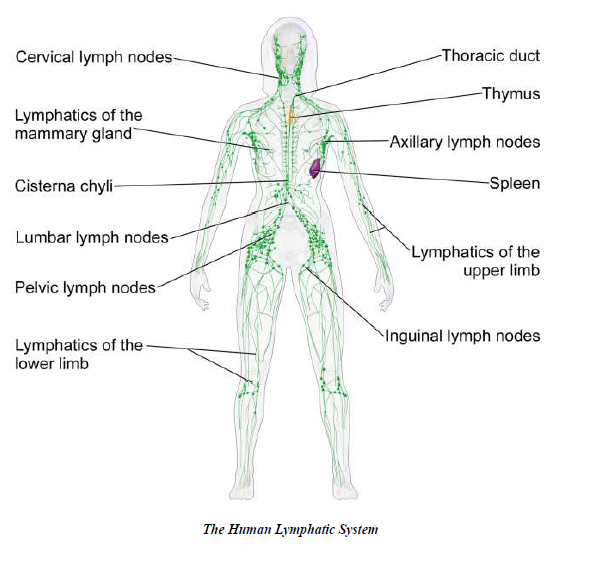
The lymphatic system is a network of tissues and organs that primarily consists of lymph vessels,lymph nodes and lymph. The tonsils, adenoids, spleen and thymus are all part of the lymphaticsystem.
There are 600 to 700 lymph nodes in the human body that filter the lymph before it returns to thecirculatory system.
The spleen, which is the largest lymphatic organ, is located on the left side of the body just abovethe kidney.
The thymus, which stores immature lymphocytes and prepares them to become active T cells, islocated in the chest just above the heart.
Tonsils are large clusters of lymphatic cells found in the pharynx.
The Components of the Human Lymphatic System
Describe the components of the human lymphatic system
The Human Lymphatic System
Functions of the lymphatic system
The lymphatic system performs the following functions:
- Removes excess fluid and waste products from the interstitial spaces between the cells andreturns it into the bloodstream.
- It also functions in transporting white blood cells to and from the lymph nodes into thebones, and antigen-presenting cells (APCs), such as dendritic cells, to the lymph nodes wherean immune response is stimulated.
- Special lymph vessels (lacteals) absorb fat and fat-soluble vitamins from the small intestineand deliver these nutrients to the cells of the body where they are used by the cells.
- Protects the body against germs. Lymph glands produce lymphocytes which produceantibodies that fight against microbes. They also contain phagocytes, which eat dead whitecells and microbes in the lymph.
The Common Disorders and Diseases of the Lymphatic System
Mention the common disorders and diseases of the lymphatic system
There are several diseases and disorders that affect the lymphatic system. The two commondisorders of the lymphatic system are lymphoedema and lymphatic filariasis (elephantiasis).
Lymphoedema
Lymphoedema is a chronic swelling of the limbs caused by the accumulation of lymph fluid thatoccurs if the lymphatic system is damaged or not functioning properly. While the limbs aretypically involved, the face, neck and abdomen may also be affected.
The lymphatic system consists of a series of lymph nodes (glands) connected by a network ofvessels, similar to blood vessels. Fluid surrounding body tissues usually drains into nearbylymph vessels so it can be transported back into the blood. However, if the lymph vessels areblocked, the fluid can’t be reabsorbed and will build up in the tissue.
Symptoms
Swelling of the limbs (arms or legs) is the common symptom of this disorder.
Effects
Unlike oedema, lymphoedema is a long-term condition that can cause discomfort, pain and a lossof mobility.
Elephantiasis
Lymphatic filariasis, commonly known as elephantiasis, is a parasitic infection that causesextreme swelling in the arms and legs. It is a painful and profoundly disfiguring disease. Whilethe infection is usually acquired in childhood, its visible manifestations occur later in life,causing temporary or permanent disability.
The disease is caused by the filarial worm, which is transmitted form human to human via thefemale mosquito when it takes a blood meal. The parasite grows into an adult worm that lives inthe lymphatic system of humans.
Symptoms
Elephantiasis is typically characterized by a thickening of the skin and subcutaneous tissue thatgives rise to the grossly enlarged and swollen limbs that earn the condition its name. In additionto the characteristic swelling, people with this disorder sometimes have bouts of fever andheadache.
Cause
The disease is caused by thread-like nematode worms, known as filariae. The larvae(microfilariae) of the parasite are taken up by the mosquito when it feeds. When the larvae reachthe third stage of development, they are introduced to a new host, who then develops theinfection.
Effects
- Filarial infection can cause lymphoedema of the limbs, genital disease (hydrocele, chylocele,and swelling of the scrotum and penis). It also causes recurrent acute attacks, which areextremely painful and are accompanied by fever.
- The infected people may have lymphatic and kidney damages.
- Sometimes the swollen limbs become infected.
- The infected person is disabled and cannot work to earn his/her living.
Causes, Symptoms, Effects and Prevention of Disorders and Diseases of the Human Lymphatic System
Explain causes, symptoms, effects and prevention of disorders and diseases of the human lymphatic system
Prevention and control
Effective treatment and preventive efforts would include:
- spraying insecticides to kill mosquitoes;
- giving antibiotics to prevent or control infection;
- giving medications to kill microfilariae circulating in the blood;
- applying pressure bandages to reduce swelling; and
- surgically removing infected tissue.
The Concept of Vascular System
Explain the concept of vascular system
TRANSPORTATION IN PLANTS
Materials to be transported across the plant body are water, minerals and food. Apart from thesenutrients, substances like the hormones also have to be transported. The transport of materialstakes place through a specialized tissue called the vascular tissue. The tissue is made up of xylemand phloem tissues. Xylem tissue transports water and mineral salts from the soil to all parts ofthe plant. Phloem tissue transports manufactured food from the sites of photosynthesis to allparts of the plant.
In between the xylem and phloem is the vascular cambium. The cells of cambium tissue divideto form a new xylem and phloem. As these cells divide and multiply, the plant increases its girth.The xylem grows inward from the vascular cambium while the phloem grows outward from thevascular cambium.
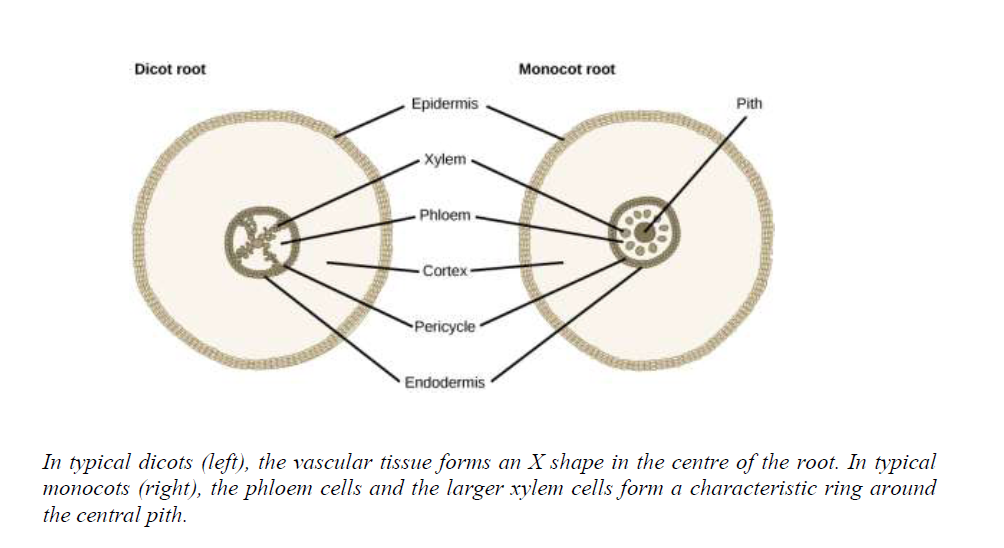
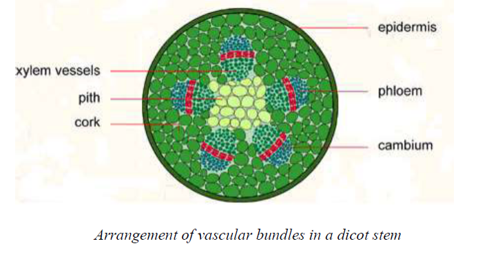
The arrangement of the vascular bundles in the stem, root and leaf of dicot and mocot plantsdiffers in a number of ways. The diagrams below show the manner they are arranged in therespective organs.
The vascular tissue in the root is arranged in the inner portion of the root, which is called thevascular cylinder. A layer of cells known as the endodermis separates the vascular tissue fromthe ground tissue in the outer portion of the root. The endodermis is exclusive to roots, andserves as a checkpoint for materials entering the root’s vascular system. A waxy substance calledsuberin is present on the walls of the endodermal cells. This waxy region, known as theCasparian strip, forces water and solutes to cross the plasma membranes of endodermal cellsinstead of slipping between the cells. This ensures that only materials required by the root passthrough the endodermis, while toxic substances and pathogens are generally excluded. Theoutermost cell layer of the root’s vascular tissue is the pericycle, an area that can give rise tolateral roots. In dicot roots, the xylem and phloem are arranged alternately in an X shape,whereas in monocot roots, the vascular tissue is arranged in a ring around the pith.In
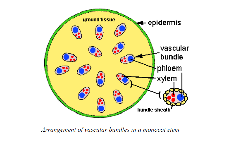
In monocot stems, the vascular bundles are scattered throughout the stem as indicated in thefigure below.
In dicot stems, the vascular bundles are arranged in a ring around the pith.
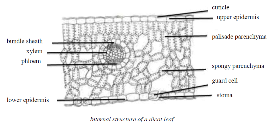
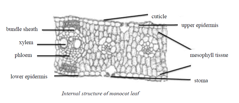
The arrangement of vascular bundles in the leaves of dicots and monocots differs. The diagramsbelow show the differences in arrangement of the bundles. Can you notice the differences? Thexylem and phloem vessels are enclosed in a bundle sheath.
Components of Vascular System
Describe components of vascular system
The vascular (transport) system in plants is made of vascular bundles. The vascular bundles aremade of xylem and phloem which are separated by a wall called vascular cambium, often simplyshortened as cambium (see diagrams discussed in the previous section).
Xylem
It is the vascular tissue that transports water across the plant body. Xylem is made up of fourdifferent types of cells. They are tracheids, vessels, xylem fibres and xylem parenchyma. Ofthese only tracheids and vessels are involved in the transport of water and minerals.
Tracheids
Tracheids are elongated dead cells that have sloping end walls. The cavity is empty as the cellsare dead. The walls are thickened with a material called lignin to prevent them from collapsingas water is transported up the plant. These thickenings are in different patterns. The cells arearranged end to end.
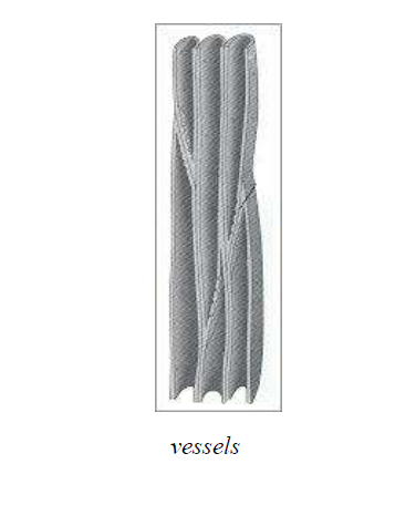
Vessels
Vessels are also dead cells that have variously patterned thickened walls. These thickenings aredue to lignin. The vessels are arranged end to end. The end walls of the vessels are eitherpartially or fully dissolved. This results in the formation of long tubes that carry water.The xylem vessels and tracheids together form long tubes that have a narrow diameter. Thus theyfunction as capillaries (narrow tubes) to transport water.
Phloem
It is the vascular tissue that transports organic substances like sucrose across the plant body. It ismade up of four types of cells namely sieve tubes, companion cells, phloem fibres and phloemparenchyma. Except for phloem fibres, all the other three types of cells are living. Sieve tubesand companion cells are mainly involved in the transport of the materials.
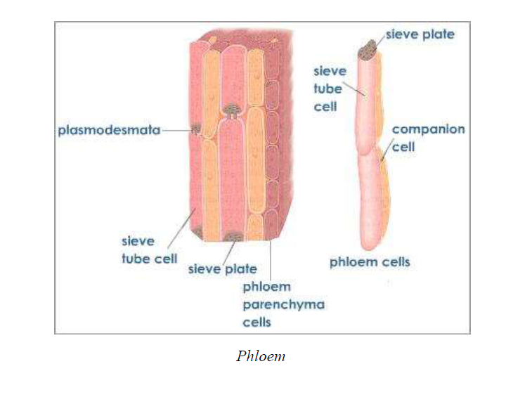
Sieve tubes
They are tubes formed by cells that are joined end to end. The end walls of these cells haveperforations. The mature sieve tube cells are enucleated. The cytoplasm of the sieve tube cells iscontinuous through the perforations of the end walls. This helps in the transport of materials.
Companion cells
They are smaller cells associated with the sieve tubes. They have dense cytoplasm and elongatednucleus. They are in contact with the sieve tube cell through pores in the wall.
The Function of Vascular System in Plants
Explain the function of vascular system in plants
The vascular system is mainly responsible for transportation of materials within a plant body.The xylem and phloem tissues are specialized to perform different functions in a plant body.
Functions of phloem
The xylem functions in transport (translocation) of manufactured food from the leaves to the cells of the plant, storage organs, fruits, etc.
Functions of xylem
- Provides support for woody plants.
- Transports water and solutes from roots to all plant parts.
The Functions of Root Hairs in Absorption and Movement of Water and Mineral Salts in Plants
Explain the functions of root hairs in absorption and movement of water and mineral salts in plants
Water and mineral uptake by roots
Plants absorb water from the soil through the root and transport it to the stem, leaves andflowers. Roots have root hairs that are unicellular, thin-walled outgrowths of the epiblema (skinof the root).
The root hairs are in close contact with the thin film of water surrounding the soil particles.There are mineral salts such as nitrates, chlorides, sulphates, phosphates, etc., dissolved in thiswater.
Water is absorbed by osmosis, while the minerals are absorbed as ions by active transport(transport against the law of diffusion, by spending cellular energy). The cell membrane hastransport proteins that allow the ions to cross the membrane. The ions then move upward throughthe xylem, to the leaves and other aerial parts of the plant.
The cell wall of each root hair is permeable to water and minerals, but its cell membrane and themembrane around the vacuole are semi permeable membranes. The root hair cells take upmineral ions by active transport.
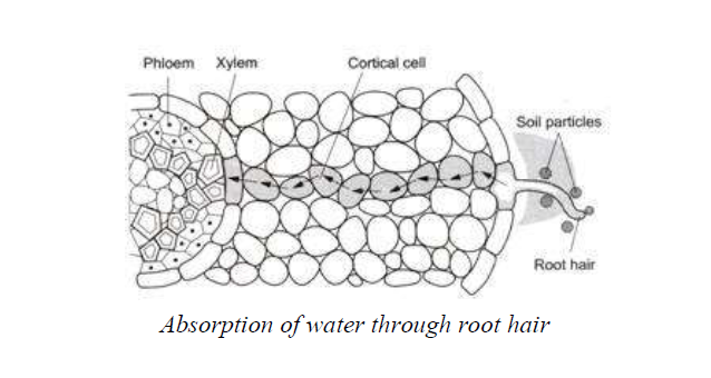
This creates a concentration difference of these ions between the root and the soil. Now, the soilsolution has higher water content than the cell sap of the root hair. Hence, water from the soildiffuses into the root hair. The root hair cells now become turgid, while the adjacent cells of thecortex have lower water content.
This results in the diffusion of water from the root hairs into the cortical cells (see figure below).After passing through the cortical cells by osmosis, the water reaches the endodermis (tissueseparating the cortex from the vascular tissues). The endodermis forces water into the xylemtubes through passage cells.
The pressure with which water is pushed into the xylem tubes of the root is called root pressure.The water moving upwards forms a column, which is maintained up to a certain height due toroot pressure.
The Movement of Water and Dissolved Mineral Salts in Plants
Outline the movement of water and dissolved mineral salts in plants
Upward movement of water within the plant
There are several processes that enable the water to move up a plant. These processes includeroot pressure, transpiration pull, cohesion, adhesion and capillarity.
Root pressure
As long as the soil is damp, there will be water taken in by the root hairs. As more water is takenin, the water that is already in the xylem vessel will be pushed up the plant. This is called rootpush or root pressure and it helps to push water up to the leaves.
Root pressure is capable, under ideal atmospheric conditions, of pushing water one or two feetabove the ground. Since root pressure is not strong enough to move water up very high, anotherprocess called transpiration pull is needed to enable the water to continue moving up the plant.
Experiments to Demonstrate Transpiration pull, Root Pressure and Capillarity
Conduct experiments to demonstrate transpiration pull, root pressure and capillarity
Transpiration pull
Transpiration is the loss of water through the leaves and other parts of the plant. Mosttranspiration occurs through openings, called stomata, on the underside of the leaves. Astranspiration occurs, water is lost. This water is replaced by water in the xylem vessels. Thiscauses an upward pull (transpiration pull or transpiration stream) on the water in the vessels.Thus, water is pulled up through the plant, and more enters by the roots to replace it.
Cohesion
Cohesion is the force of attraction between similar molecules. Transpiration pull is possiblebecause water molecules cling to each other by cohesion. When water molecules cling to eachother as they move up the stem and into the leaves, they pull up more water molecules up theplant. This process, however, is facilitated by transpiration pull since the water molecules lostthrough transpiration is being replaced by more water molecules absorbed by the roots.
Adhesion
Adhesion
Adhesion is the force of attraction between different molecules. As water molecules are stucktogether by cohesion, the entire column of water in the xylem adheres to the sides of the xylem.It is said that the water in under tension as the column moves up the xylem. At the same time, thexylem tube narrows because of the tension.
Cohesion and adhesion forces maintain a continuous column of water in the xylem vessels fromthe roots to the leaves of plants.
Capillarity
Capillarity is the tendency of water to rise through narrow tubes. The lumen of xylem tracheidsand vessels is very narrow and this enables water to rise through it by capillarity. Capillarity isassisted by adhesion and cohesion forces.
The Concept of Transpiration
Explain the concept of transpiration
Transpiration is the evaporation of water from plants. It occurs chiefly through the leaves whiletheir stomata are open for the passage of carbon dioxide and oxygen during photosynthesis.
Transpiration also occurs through the cuticle and lenticels. Lenticels are pores in the stems ofwoody plants that allow gaseous exchange between the atmosphere and the internal tissues.
The Significance of Transpiration in Plants
Outline the significance of transpiration in plants
Transpiration is of immense importance in plant life as it is of great benefit to the plant. Thefollowing are the reasons why transpiration is important in plants.
Cooling of the plant
The leaves absorb the radiant energy. Some of the light energy is utilized in photosynthesis. Therest is converted into heat energy resulting in an increase in leaf temperature. However, rapidloss of water in the form of water vapour from the aerial parts of the plant through transpirationbrings down their temperature. Transpiration thus provides a significant cooling effect whichkeeps the plant from being overheated.
Mineral transport
Mineral salts remain dissolved in the soil water and are absorbed by the roots. Minerals that arcabsorbed and accumulated in the xylem duct of the root move up and are distributed in the plantby the transpiration stream.
Water movement
The absorbed water is transported from roots to leaves through the xylem vessels. This is greatlyinfluenced by transpiration pull. Water loss due to transpiration results in the development of lowwater potential in the leaf tissues. Thus, water moves from the xylem vessels to the leaf cells.
Development of mechanical tissues
Greater amount of transpiration helps in the development of mechanical tissues in plants. Theplants become healthier and more compact, the cell walls become thick and cutinized and theplants are able to resist the attack of fungi and bacteria.
Maintenance of turgidity
Transpiration maintains an optimum degree of turgidity in cells. Under favourable conditions,plants absorb excess amount of water, which is given off by transpiration to maintain theoptimum turgidity for better growth.
Increase of taste of fruits
The solutes inside the cell become more concentrated when transpiration is rapid. Consequently,the concentration of sugar solution in the cells of fruits increases and fruits taste sweeter.
Wilting
When the rate of evaporation is higher than that of absorption of water from the soil, as it occursduring drought conditions, the plant wilts. Wilting is beneficial when a plant cannot obtainenough water to replace that lost by the plant through transpiration because it causes the closureof the stomata (singular: stoma). Thus, the rate of evaporation is greatly reduced.
Transpiration as a necessary evil
Transpiration is a necessary evil because of the following facts:-
- A large amount of absorbed water is lost during transpiration which is harmful to plants.
- Unnecessary wastage of energy takes place during the process of water absorption which islost due to transpiration.
- When the rate of transpiration is high in plants growing in soil deficient in water, an internalwater deficit develops in plants which may affect metabolic process.
- Many xerophytes undergo structural modifications and adaptations to check transpiration.
Considering both the beneficial and harmful effects of transpiration, it may be concluded that itis definitely advantageous in spite of its harmful consequences.
Factors Affecting the Rate of Transpiration in Plants
Outline factors affecting the rate of transpiration in plants
The rate of transpiration can be affected by both plant features and environmental factors.
Plant factors
These plant parameters help plants control rates of transpiration by serving as forms of resistanceto water movement out of the plant. They include the following:-
Root system
Plants with extensive root systems absorb a great amount of water and therefore much water ismoved up the plant. Thus, plants with extensive root systems have higher rates of transpirationthan those with few roots.
Size of leaves
A plant with broad leaves tend to lose more water than that with small leaves. This is because thebroad leaves have large surface areas over which transpiration takes place.
Leaf structure
The structure of a leaf has a great influence on the rate of transpiration. The following areanatomical structures of a leaf that affect the rate of transpiration:-
Number of stomata
Stomata are pores in the leaf that allow gaseous exchange to take place, and water vapour toleave the plant. Special cells called guard cells control each pore’s opening or closing. Someplants have many stomata while others have a few stomata. The more the stomata, the higher therate of transpiration and vice versa.
Position of stomata
Plants with few stomata on the upper surface of the leaf experiences a little transpirationcompared to those with many stomata on the lower leaf surface. This is because the uppersurface is highly stricken by direct sunlight hence increasing the rate of transpiration.
Epidermal hairs
Epidermal hair on the leaf traps a thin layer of still air close to the leaf surface. For the water lostfrom the leaf to get into the atmosphere, it has to cross this resistant layer of air. The layer thuschecks excessive loss of water from the leaf. Likewise, the water vapour from the leaf is alsotrapped by the epidermal hairs. This prevents further loss of water vapour from the leaves andhence slows down the rate of transpiration.
Size of stomatal air spaces
Large air spaces between the cells of the spongy mesophyll and stomata, called substomatal airspaces, increase the rate of transpiration. Small substomatal air spaces reduce the rate oftranspiration.
Cuticle
The cuticle is the waxy layer present on all above-ground tissue of a plant and serves as a barrierto water movement out of a leaf. Because the cuticle is made of wax, it is very hydrophobic or‘water-repelling’. Therefore, water does not move through it very easily. The thicker the cuticlelayer on a leaf surface, the slower the transpiration rate. Cuticle thickness varies widely amongplant species. In general, plants from hot, dry climates have thicker cuticles than plants fromcool, moist climates. In addition, leaves that develop under direct sunlight will have muchthicker cuticles than leaves that develop under shade conditions.
Environmental factors
Some environmental conditions create the driving force for movement of water out of the plant.Others alter the plant’s ability to control water loss.
Light
Plants transpire more rapidly in the light than in the dark. This is largely because light stimulatesthe opening of the stomata (mechanism). Light also speeds up transpiration by warming the leaf.
Photosynthesis occurs in the presence of light. A higher light intensity increases the rate ofphotosynthesis in the guard cells. As the guard cells absorb water from the soil forphotosynthesis, they become turgid and hence the stomata are opened, and hence a higher rate oftranspiration.
Temperature
Plants transpire more rapidly at higher temperatures because water evaporates more rapidly asthe temperature rises. At 30°C, a leaf may transpire three times as fast as it does at 20°C.
Relative humidity
Relative humidity is the amount of water vapour in the air compared to the amount of watervapour that air could hold at a given temperature. When the air is less moist, the relativehumidity is low, and thus the rate of transpiration is greater. When relative humidity is high, theatmosphere contains more moisture, reducing the rate of transpiration. Therefore, transpirationincreases with the decrease in relative humidity.
The rate of diffusion of any substance increases as the difference in concentration of thesubstances in the two regions increases. When the surrounding air is less humidity, diffusion ofwater out of the leaf goes on more rapidly.
Wind
The wind removes water vapour and thus increases the rate of transpiration. High winds lead tostomatal closure to stop the rapid water loss and hence bring a drop in rate of transpiration.Moderate winds may reduce transpiration by lowering the temperature of the leaf.
When there is no breeze, the air surrounding a leaf becomes increasingly humid thus reducingthe rate of transpiration. When a breeze is present, the humid air is carried away and replaced bydrier air, thus increasing the rate of transpiration.
Soil water
The source of water for transpiration out of the plant comes from the soil. Plants with adequatesoil moisture will normally transpire at high rates because the soil provides the water to movethrough the plant. Plants cannot continue to transpire without wilting if the soil is very drybecause the water in the xylem that moves out through the leaves is not being replaced by the soil water. Thus, the rate of transpiration will increase when there is adequate amount of water inthe soil and will decrease when the soil contains little mositure.
Atmospheric pressure
Transpiration is high at low atmospheric pressure and it is low at high atmospheric pressure.Plants that grow naturally at higher altitudes, where the atmospheric pressure is low, havemodified leaves to reduce the rate of transpiration.
- READ TOPIC 5: Gaseous Exchange And Respiration